Platforms
Five discovery routes, one team
Running all five means we can recommend against a platform without recommending against ourselves.
Which one fits your antigen?
| Transgenic mouse | When the program is heading toward IND filing and you want fully human antibodies from day one. Needs an antigen suitable for immunization. |
|---|---|
| Hybridoma | When you need reliable monoclonals as research tools, diagnostic reagents or structural supply, or a diverse multi-epitope panel. |
| Single-B cell cloning | When native VH/VL pairing matters, the response is low-frequency, or you are mining a convalescent or immunized repertoire. |
| Yeast display | When you already have a lead and need higher affinity, or need to screen for stability and expression before committing. |
| Phage display | When the target is hard to immunize against — GPCRs, ion channels, haptens, conformational epitopes — or you need VHH formats. |
Platform 01
Human IgG Transgenic Mouse
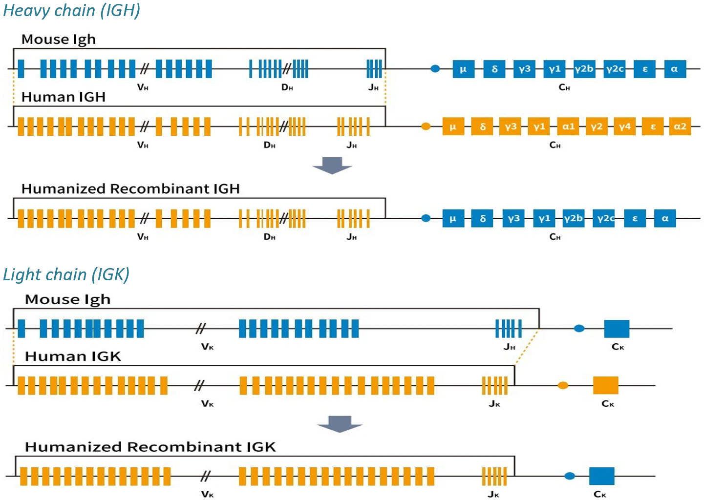
Variable regions are encoded by humanized Igh and Igk loci, so the antibodies that come out are already fully human. That removes humanization from the program entirely, along with its cost and timeline.
Because selection happens through the animal's own germinal center reaction, leads arrive already affinity matured in vivo. A fully human framework also carries lower immunogenicity risk, and in practice gives cleaner SEC profiles than chimerized leads.
Not the right route for antigens that cannot survive immunization — use phage display instead.
Platform 02
Hybridoma
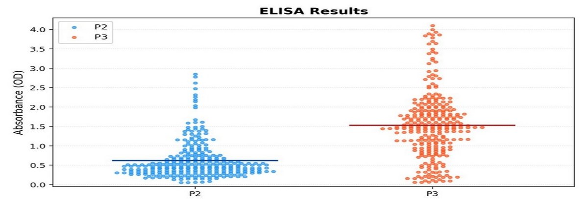
A standard campaign runs four mice with three bleeds each, tracked by serum titer before fusion. Around 1,000 clones are screened by ELISA, positives amplified by PCR and confirmed by Sanger sequencing.
Typical outcomes: roughly 70% of clones give OD450 above twice background, with sequence clarity above 95% on confirmed clones.
Still the workhorse for research-tool monoclonals, companion diagnostic antibodies, and consistent supply across long structural biology projects. Also the natural route to a diverse multi-epitope panel — the raw material for epitope binning and finding sandwich pairs.

Platform 03
Single-B Cell Cloning
Fusion-based methods lose clones, and what survives is biased by which cells fuse well rather than which antibodies are best. Sorting single B cells by FACS and amplifying heavy and light chain from each cell individually preserves the natural pairing and sidesteps that loss.
This is the route to high-diversity responses from immunized animals or convalescent donors, where the interesting antibodies may be present at low frequency. With no fusion, outgrowth and subcloning sequence, a diverse panel also arrives appreciably faster.
Platform 04
Yeast Display
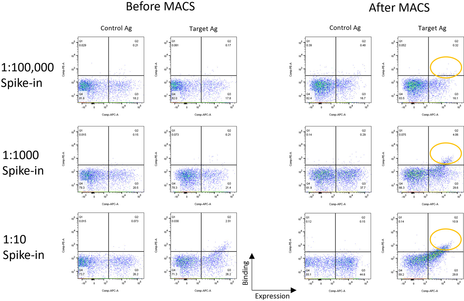
Validated recovery of rare antigen-specific clones across three dilution ratios — 1:10, 1:1,000 and 1:100,000. Three rounds of MACS consistently enrich the target-binding population, giving tight FACS populations afterwards.
The natural home for affinity maturation: start from a naive library, or build an error-prone PCR library from an existing hit and sort for improved variants. Because display level correlates with expression and stability, it also allows selection for developability alongside affinity.
The same platform serves enzyme engineering, directed evolution, and protein interaction studies — groups come to us for this without any antibody program attached.
Platform 05
Phage Display
We build target-specific libraries optimized for your antigen rather than panning a generic library and hoping. This matters most for difficult-to-immunize targets — GPCRs, ion channels, haptens and conformational epitopes — where immunization often produces nothing usable.
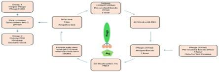
Display also lets you control selection conditions. We can select under physiological or stress conditions — pH, temperature, ionic strength — so binders are chosen for tractability as well as affinity. Selection runs three to four rounds against antigen-coated beads, with hits confirmed by phage ELISA and colony PCR.
Custom scFv, Fab and VHH libraries. Single-domain formats open epitopes conventional antibodies cannot reach.